During the Francophone Diabetes Society convention, a special plenary session was held on diabetic neuropathy. This problem, which is currently widespread, is growing more widespread and is not being sufficiently addressed. It is imperative to phenotype patients, and neurofilament is not a suitable tool for this task. According to Agnès Hartemann, MD, PhD, head of the diabetology department at Pitié-Salpêtrière Hospital in Paris, France, electromyography (EMG) can also be deceiving.
According to Liane Ong, PhD, lead research scientist at the University of Washington in Seattle and coauthor of the 2021 Global Burden of Disease, Injuries, and Risk Factors Study, “the number of people affected by diabetic neuropathy has more than tripled worldwide since 1990, reaching 206 million in 2021.”
The literature indicates that neuropathic pain affects 25–30% of those with diabetic peripheral neuropathy. Age-related pain progression is not surprising. In the 26-year observational follow-up of the DCCT trial (1982–1993), which followed the Epidemiology of Diabetes Interventions and Complications study, the prevalence of neuropathic pain (as measured by the answers to the questions “Have you ever felt burning sensations in your legs and/or feet?” and/or “Does it hurt when bed covers touch your skin?”) rose from 8.5% to 19.8%. From 22.9% to 43.5%, the percentage of people have a Michigan Neuropathy Screening Instrument score greater than 2.
“The monofilament has taken an excessive place in screening,” commented Hartemann during his congress presentation on the etiology of neuropathic pain. It has long been thought that large fibers were impacted by painful neuropathy and tiny fibers by sensory neuropathy. This difference is no longer valid, though, as both kinds of fibers may be involved in different forms of neuropathy.” Peripheral neuropathy really manifests in two different ways: first, as “sensory” neuropathy, where there is a loss of function due to fiber loss; second, as hyperexcitability, or hyperactivity of fibers, which results in a gain of function. This hyperactivity is caused by malfunctioning ion channels that activate spontaneously, repeatedly, and prematurely at the peripheral level, causing problems at the spinal junctions.
Gain and Loss of Function
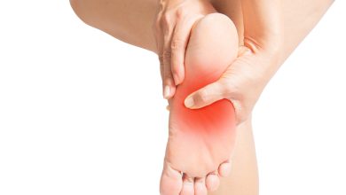
A rarefaction of both large (> 30 m/s) and tiny (3-30 m/s for thinly myelinated and < 3 m/s for unmyelinated) nerve fibers is associated with the loss of function, or sensory neuropathy. “When you look for this loss of function in large fibers, that’s where you’ll find the abolition of osteotendinous reflexes, decreased vibration and proprioception perception, sensitivity to touch and pressure,” Hartemann stated. The 10-g monofilament test investigates the following: a gentle press and touch interaction. Reduced pain sensitivity (as measured by the needle test), temperature and pressure sensitivity, and feeling of heat and cold are caused by the rarefaction of small fibers, and these effects appear to be common to both big and small fibers.
Furthermore, Hartemann noted that hyperexcitability “can come from large fibers,” proving that painful neuropathy (loss of function) affects more than simply small fibers as previously believed. Patients thus experience mechanical allodynia (rubbing of sheets or cotton) and a feeling as though the foot is gripped in a vise. The well-known symptoms of prickling, burning, itching, thermal allodynia, hyperalgesia, severe cold (the sensation of walking barefoot on snow), and electric shocks are caused by hyperexcitability of the tiny fibers.
There is a lack of inhibitory pain function in the spine. Increased anxiety, despair, and pain-related sleep problems are among the brain’s consequences of hyperexcitability. However, these disruptions are more frequent and last longer than those seen in chronic pain of comparable severity but different origin, with a vicious cycle involving the peripheral, spinal, and central nervous systems amplifying the effects.
It’s unknown if neuropathy starts with loss of function or hyperactivity of the fibers. The population and the instruments employed determine the proportion of patients who exhibit one neuropathy, the other, or both. Researchers discovered deafferentation in 54% of 232 patients (74%), “irritable nociceptors” in 15%, and both in 31% of patients with neuropathy verified by EMG or biopsy. The patients had an average age of 63 years.
EMG When Uncertain
Fiber rarefaction, often known as sensory neuropathy, is primarily diagnosed clinically. Only if big fibers are affected by the loss of function might EMG show abnormalities. Therefore, even while there is targeted involvement of tiny fibers, it is feasible to mistakenly assume that diabetic neuropathy does not exist in the absence of EMG anomalies.
Clinical research uses skin biopsy at the ankle, which reveals rarefaction of tiny fibers in the dermis and epidermis, to phenotype individuals. There is currently no standard for confocal corneal microscopy, which is the indirect vision of tiny fiber loss.
Excitation, or hyperactivity, can also be diagnosed clinically. For a positive diagnosis, EMG, skin biopsy, and confocal corneal microscopy may be normal. “We must refer our patients to pain centers so that they are phenotyped and receive the most appropriate treatment for the type of pain,” Hartemann stated. Diabetic neuropathy must be identified, especially in people with the disease, as they may experience a variety of aches, particularly in the lower limbs.
Several diabetic neuropathy teams have revalidated the DN4 screening questionnaire for this reason. With an 83% sensitivity and 90% specificity, diabetic neuropathy is suggested by a score greater than 4.
According to a 2013 study in which Hartmann took part, diabetic neuropathy affected 14% of individuals with type 1 diabetes and 24% of patients with type 2 diabetes. Of those who sought medical advice for pain, almost 70% had done so, but only 38% had gotten the right care.
A neurologist should be consulted if any of the following traits—such as the rapid onset, symmetry, significant motor loss, or proximal involvement—raise questions regarding the diagnosis of diabetic neuropathy.
Cervical, dorsal, and lumbar radiculopathy are examples of concomitant diagnosis. Both MRI and EMG are pertinent in these situations. Peripheral artery disease, osteoarthritis in the knee, Parkinson’s disease, post-stroke neuropathy, and chemotherapy are further etiologies to take into account.
Microangiopathy, which is caused by damage to the microvessels that innervate the nerves, is a characteristic of neuropathy. However, there are other risk factors for neuropathy, such as smoking, dyslipidemia, hypertension, obesity, metabolic syndrome, and glycemia. According to Hartemann, “it even starts in type 2 prediabetes.” Thus, axonal insulin resistance and chronic hyperglycemia (microangiopathy on endoneurial capillaries) are associated with the same risk variables as muscle. There is endoplasmic reticulum stress, oxidative stress, and malfunction of the axon mitochondria.